The international congress of forensic-medico-legal imaging, organized by the University of Toulouse 3 Paul Sabatier and the International society for forensic radiology and imaging (Isfri), which takes place from May 25 to 27 at the Paladia hotel in Toulouse, will bring together 200 practitioners. Interview with one of the pioneers of the virtual autopsy, the radiologist and pathologist of the CHU of Toulouse Fabrice Dédouit.
The international congress of medical imaging, organized by the University of Toulouse 3 Paul Sabatier and the International society for forensic radiology and imaging (Isfri), which takes place over three days (May 25-27), highlights the new technologies in forensic medicine?
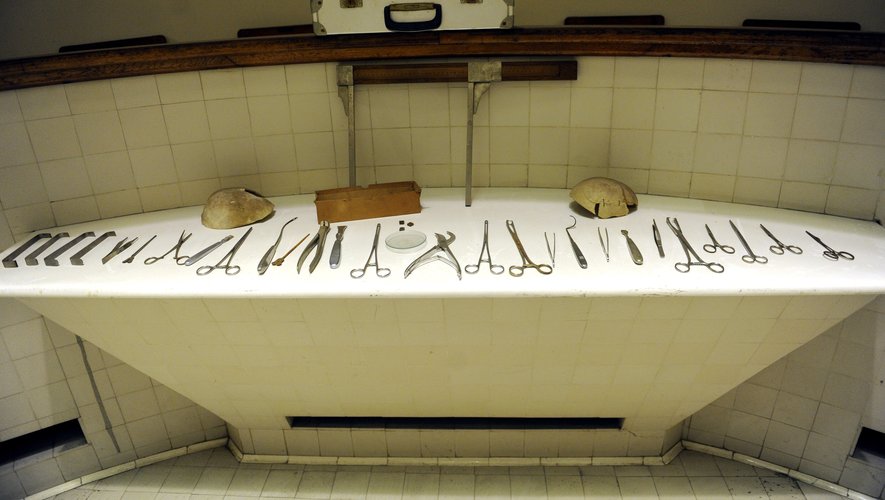
Among the themes addressed, there is indeed the virtual autopsy or virtopsy. The virtual autopsy uses modern cross-sectional imaging, mainly MRI or CT on cadavers. It is practiced before the autopsy and it allows, depending on the indications, to help the medical examiner. It does not replace the autopsy. We are not in a philosophy of substitution, but in a philosophy of complementarity. It is therefore an additional examination. During an autopsy, the doctor may have to take several samples which will be analyzed later in order to help determine the causes of death. Among all these complementary techniques, there is of course imaging, but also toxicology, genetics, chemistry, limnology for drowning diagnoses, etc. All these additional examinations will help the medical examiner to establish his diagnosis.
What about virtopsia, a technique presented as a revolution supposed to reveal the secrets of death?
It is the virtual autopsy performed on the corpse. The classic forensic autopsy is smart. If we have new tools to move forward and answer the questions asked by magistrates and investigators, we use them, it’s complementarity.
The method of virtopsia is characterized in particular by the injection of a contrast product which makes the blood vessels of the deceased more visible?
There are several techniques: CT scan without injection of product and post-mortem CT angiography where a contrast product is injected into the corpse. This requires first having vascular, arterial and venous access. The most widely used contrast products in the living are those that are iodized. This allows the vessels to be filled in order to obtain a contrast of the entire vascular system. For the corpse, it is a specific contrast product and a specific injector. But it’s the same principle in the clinic as the injected scanner, with perfusion, a tubing connected to an injector filled with iodine. With a corpse, there are peculiarities, the heart does not beat and there is no circulation. Virtopsia is a method that was developed in Switzerland about fifteen years ago. There are very few hospitals in France that are equipped with the machine. In Toulouse, we have this system at the CHU.
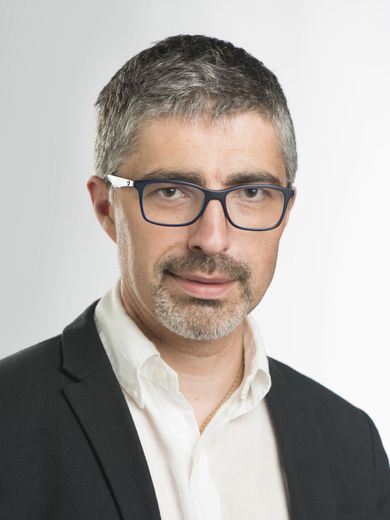
Fabrice dédouit, radiologist and medical examiner at the University Hospital of Toulouse.
Has virtopsia become systematic during an autopsy?
No not at all. It depends on the case, the context, the opinion of the magistrate, the medical examiner. From the moment there is a medico-legal obstacle that has been signed by a doctor, the body of the deceased no longer belongs to the family. Several options are possible: either we do a simple external examination, that is to say that we do not open the body, we undress it and describe the lesions; or there are doubts and we practice virtopsia. Which is a preparatory time for the autopsy. Depending on the case, there may be additional procedures.
This method is supposed to provide elements of precision. Is this the purpose of this congress on forensic imaging which brings together nearly 200 forensic doctors, dentists, anthropologists and scientists from 24 countries?
Progress is related to technology. It’s like in the clinic. what is interesting is that concerning the corpse, we are not bound to the radiation protection measures, which are on the other hand the constraints for the living. And for children in pediatrics, it’s even more restrictive because of the rays that we keep for a lifetime. On the corpse, there is no such limit. Sub-millimeter spatial resolutions and exceptional image quality are therefore obtained. We see vessels that we do not see during a CT scan injected into the living. It’s truly revolutionary. Things in France are progressing very well. Personally, I’ve been using forensic imaging for twenty years and I can’t do without it.