A Toulouse start-up has developed an algorithm that can more quickly detect biomarkers of epileptic seizures, but also those of the epileptogenic focus. A breakthrough thanks to artificial intelligence that gives hope for improving surgical procedures
The crises are impressive. And very trying for those who suffer them. Epilepsy is the third most common neurological disease in France and affects 500,000 people in the territory, or 1% of the population of France. If some have only one or two attacks in their childhood, and never have them again afterwards, other patients will have them very regularly throughout their lives. To the point of becoming a real handicap.
30% of operated patients still have seizures after
To treat this pathology, drug treatments exist. And 60 to 70% of patients are receptive to it. But for the others, nearly 5,000 people in France, “the only solution, if the drug treatment does not work, is to remove by surgery the part of the brain that generates epileptic seizures and that we called the epileptogenic zone. In France, less than 500 patients undergo this type of surgery each year,” says Ludovic Gardy, CNRS research engineer at the Brain and Cognition Research Center (CerCo). He conducted a doctoral thesis on the subject, straddling ENAC and CerCo, and co-founded the Toulouse start-up Avrio MedTech.
[#CNRSInnovation] The tool to precisely identify the epileptogenic focus using innovative biomarkers will be developed at the heart of the startup #AvrioMedTecha subsidiary of the CNRS.
Works by Ludovic Gardy and Emmanuel Barbeau from #CerCo, a unit affiliated with the MSHS-T. https://t.co/caUsuwzuDk— MSHS TOULOUSE (@mshstoulouse) May 12, 2023
The latter is working on a new device capable, thanks to artificial intelligence, of better identifying the epileptogenic focus, the area at the origin of the seizures, because, in 30% of cases, operated patients still have seizures after surgery. surgery due to poor targeting.
To remove this part of the brain, it is necessary to locate it to the nearest millimeter. To delimit it, the patients initially undergo a battery of examinations, from MRI to electroencephalography (EEG) of the scalp which records the cerebral signal. Sometimes, it is necessary to go further and an intracerebral exploration is necessary.
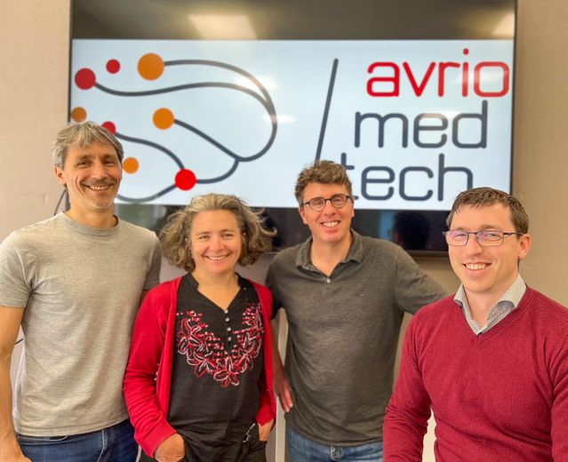
The 4 co-founders of AvrioMedTech, Christophe Hurter, Professor at ENAC, Karine Seymour, future CEO of Avrio MedTech, Emmanuel Barbeau from CNRS and Ludovic Gardy from Avrio Med Tech.
To achieve this, a neurosurgeon implants about ten electrodes, rods about ten centimeters and less than one millimeter in diameter, each capable of recording neuronal activity through various channels. If we multiply the number of channels by that of the number of electrodes, we arrive at a hundred recordings distributed in the brain of the patient. Data recorded 24 hours a day, for the entire duration of the patient’s hospitalization, ie 10 to 15 days.
Read also :
Hearing loss, epilepsy: early signs of Parkinson’s?
“What makes the amount of data to be analyzed by the epileptologist is colossal, it’s as if I asked you to read a 500-page novel every day, to give me a summary and to highlight words inside, which in this case are not words but biomarkers of epilepsy. This is obviously not possible, ”notes Ludovic Gardy.
Specific biomarkers
“We have created an automatic detector, therefore an algorithm, of pathophysiological oscillations in epilepsy. It is also able to detect a new type of biomarker specific to the epileptogenic zone, it is only found there, almost in real time. Epileptologists cannot see it because it is invisible to the naked eye, it is a kind of extremely rapid oscillation which lasts only a few thousandths of a second called fast ripples “, continues the researcher. These tangible results will help the epileptologist to identify the area concerned. Until now it was necessary to wait several weeks, whereas with the algorithm developed by Avrio MedTech, it is possible to have them almost instantly, while the patient is in the hospital.
“This result will not tell the doctor what to do, but it will provide him with extremely important elements of response in his reflection and his diagnosis and the doctor will make his information converge with all the other information he has. its disposition on the clinical case of the patient, on his history, on the other MRI imaging sections”, insists Ludovic Gardy.
Read also :
REPORTING. Epilepsy: how to live with the disease
Having lifted the brake on the ultra-rapid detection of these fastripples,and automated data management, the Toulouse start-up conducted a study on more than 50 patients in total to confirm that the results were consistent with what was expected. “All the information converges on the significant usefulness of these rapid oscillations in the prognosis. This remains at the research stage, these oscillations have never yet been used in the context of the clinic and this will be the subject of a study which starts this year and which will last 3 or 5 years, where we will clinically use this information delivered by our solution to create pools of patients who will benefit from the solution, versus others who will not benefit from it, and we will see at the end if those who have benefited from it have a better prognosis than those who have not benefited from it”, explains the co-founder of Avrio MedTech. The study will start this summer and should last three to five years. In the meantime, nothing prevents research teams from using this new tool.