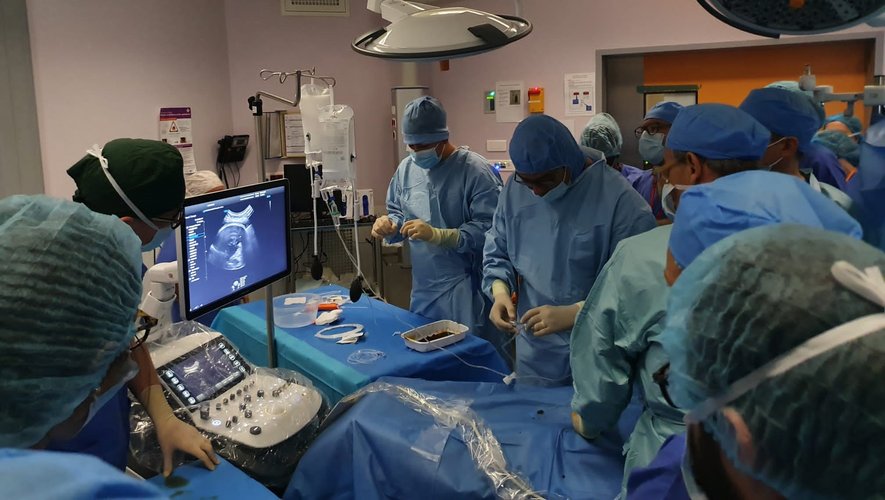
He was still in his mother’s womb when an AP-HP team operated on him in September for an aneurysmal malformation of Galen’s vein. Update on this world premiere.
An exceptional operation. A 33-week-old fetus was operated on for an aneurysm in utero, on September 7, 2022, at the Necker Hospital – Children Sick in Paris. The baby was born on October 16. Eight months after his birth, this Friday, June 16, the AP-HP announces his final cure. He does not suffer from any neurological or cardiac sequelae and is developing completely normally.
World premiere?The teams of @hospital_necker And @GhuParis treated by in utero embolization an aneurysmal malformation of the vein of Galen on 07/09/2022. The child was born on 10/16/2022 and is doing well with no sequelae.https://t.co/4Y2zRDHPYz pic.twitter.com/o0AyWGL17z
— AP-HP (@APHP) June 16, 2023
The fetus suffered from an aneurysmal malformation of the Vein of Galen (AVGM), a congenital vascular malformation in the central part of the brain. Visible on ultrasound in the second or third trimester, this rare malformation is characterized by abnormal communication between the arteries and the vein of Galen.
Serious neurological sequelae
AVVM “can have serious consequences on the development of the brain and can lead to death at birth. After this period, serious neurological sequelae and developmental delays are frequent”, notes the AP-HP in a press release. “In babies who suffer from it, we can also observe an abnormal increase in the cranial circumference due to excess cerebrospinal fluid, heart failure and difficulty in breathing (the heart being overstretched and saturated due to the very high flow rate in the malformation )”, adds the Vaud university hospital center.
Following an antenatal discovery of AGVM, the options so far have been bleak and restricted, boiling down to “the possibility of a therapeutic interruption of pregnancy or, at birth, depending on the case, a limitation of care in the event of irreversible brain damage or neuroradiology treatment by embolization”, details the AP-HP.
A multidisciplinary team
The fetus operated on in September already had heart problems but were reversible and a brain which appeared normal on the MRI. But “the risk of brain damage developing in utero until the end of pregnancy, the very large dimensions of the malformation carried a 90% risk of failure and/or death at birth”. It is therefore the fetal intervention in utero which was proposed to the family, who, informed of the risks, accepted.
A team made up of gynecological surgeons and interventional neuroradiologists treated the fetus by embolization, a procedure which consists of obstructing a blood vessel. More concretely, “Under ultrasound guidance, a microcatheter was positioned in Galen’s vein, through the mother’s skin and uterus, then through the fetal skull, allowing the deployment of 5 meters of platinum wire (coils) up to ‘to obtain a marked slowing down of the malformation’, details the AP-HP. The operation lasted 30 minutes and caused no complications.
The same operation carried out since in Boston
The pregnancy continued without difficulty, with “a marked improvement” fetal heart functions and “a reduction in the dimensions of the veins involved in the malformation measured by MRI“. Complementary embolizations were then performed on 5e and 67e baby’s day of life. He is now saved. For the doctors of the AP-HP, “Antenatal intervention may prevent, in some high-risk fetuses, the onset of serious cardiac and cerebral consequences during the third trimester and death at birth”.
Since then, the same intervention has already been performed in a Boston hospital during the first trimester. The two teams, American and French, were in close contact in order to best prepare for this remarkable intervention.

